
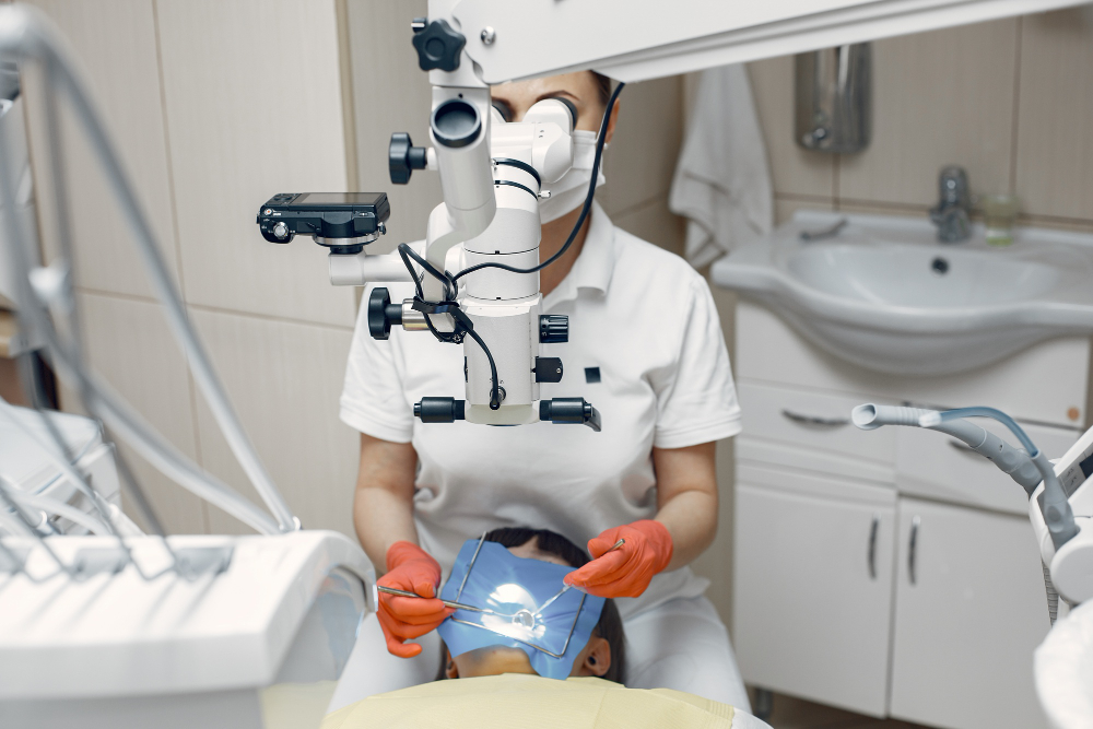
What Is a Root Canal Under Microscope?
Many people feel nervous when they hear the words “root canal.” However, new technology is changing this experience. A root canal under microscope uses a special dental microscope. This tool helps dentists see tiny details inside your tooth. Because of this, the dentist can work with much greater accuracy. In many cities, you can now find clinics offering microscope-assisted root canal treatments. This modern root canal procedure is part of precision dentistry, which aims to make dental care safer and more effective for everyone.
How Microscope Technology Improves Root Canal Procedures
Microscope technology has changed the way dentists perform root canals. First, the microscope gives a clear, enlarged view of the tooth’s inside. As a result, dentists can spot small cracks or hidden canals that are easy to miss with the naked eye. In addition, the bright light from the microscope helps dentists see even the tiniest details. Because of these features, advanced dental technology makes the treatment more precise and less likely to miss infection. According to the Journal of Endodontics, microscope use can increase the success rate of root canal therapy (1).
Benefits of Microscope-Assisted Root Canals for Patients
Choosing a microscope-assisted root canal offers many advantages. For example, patients often experience:
Furthermore, precision dentistry helps reduce the chance of future problems. Because the dentist can see so well, they can remove all infection and seal the tooth properly. This leads to a healthier mouth and a faster recovery.
Step-by-Step: What to Expect During the Procedure
Knowing what happens during a modern root canal procedure can help you feel calm. Here is what you can expect:
Throughout the process, the microscope helps the dentist work with great care. In many dental offices, you can even ask to see the microscope in action.
Safety and Success Rates: What the Research Shows
Many studies show that microscope-assisted root canal treatments are very safe. For instance, research in the International Endodontic Journal found that using a microscope can increase the success rate of root canals by up to 10% (2). Additionally, the risk of missing a hidden canal or leaving infection behind is much lower. Because the dentist can see so clearly, they can avoid mistakes and protect healthy tooth tissue. The World Health Organization also supports the use of advanced dental technology to improve patient safety and care (3).
Aftercare and Recovery Tips
After your root canal under microscope, proper care helps your tooth heal well. Here are some simple tips:
Most people recover quickly from a microscope-assisted root canal. Because the treatment is so precise, healing is often faster and more comfortable.